Eye Fundus Control for diabetic and hypertensive
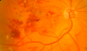
Diabetes and hypertension are chronic diseases that cause significant damage especially at the level of the vasculature (arteries and veins) in the retina of the eye.
Such damages can cause loss of vision secondary to bleeding inside the eye, inflammation of the retina, retinal detachment, increased eye pressure (glaucoma) and cataracts. Some of these conditions such as cataracts are reversible without leaving permanent visual damage, however others such as retinal detachment and glaucoma can cause irreparable loss of vision.
To avoid this it is necessary an early detection of diabetic or hypertensive damage and consequently quick treatment without delay as soon as relevant. We recommend that anyone who is a carrier of diabetes and hypertension have a ophthalmological control including an eye fundus control at least once a year, and more frequently if there is already significant damage.
Refraction (measure of glasses)
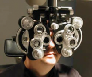 The focus of the eye defects is very common, everyone at some point have heard words like myopia, farsightedness or astigmatism. In fact almost everyone has some degree of them, although most are too small to significantly interfere with their vision.
Myopia is characterized in that the eye has too much focusing power, the affected person looks good for short distances, such as reading a book, or thread a needle, but sees blurry half or long distances. Cannot see the signs on the street, interpret traffic signs or to recognize a person until having it in a short distance. Meanwhile farsightedness causes blurred vision at all distances because the eye lacks power to focus light well; in astigmatism focus is irregular and can coexist with any of the other two entities in varying degrees. Refraction consists in the quantification of these focus defects and once these measures are taken is possible to prescript the best suited glasses or contact lenses for the patient.
Glaucoma Control and Intraocular Pressure Check
 Glaucoma is another disease that is necessary to mention due to its importance. After age 40 one in 100 people develop glaucoma without realizing it and this increases with age. The eye contains structures as crystalline, mentioned above, and transparent fluids that fill it, give its shape and consistency. These elements generate pressure on the inner walls of the eye known as the intraocular pressure. In glaucoma there is a chronic and persistent elevation of this pressure, usually by defects in the systems output eye fluid. This sustained hypertension generates damages on the nerve of the eye or optic nerve. The same works by transmitting information between the eye and the brain so that this one can interpret to be capable of seeing. When the nerve is damaged vision deteriorates, unfortunately this process is irreversible and the nerve damage cannot be repaired. To detect the disease it is required a detailed and complete eye examination including intraocular pressure exam and revision of the nerve of the eye, sometimes this is not enough and it is necessary to do some complementary tests that explore the role of the nerve of the eye, the corneal thickness and also that detects loss of nerve communications within the eye.
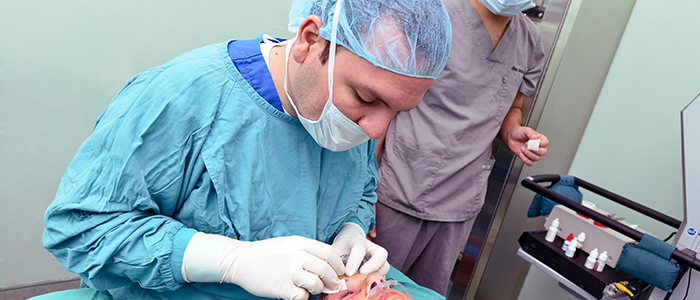
Fortunately we have effective treatments for this disease, with which in most cases control the progression of damage and avoided blindness. In patients in whom medical therapy is insufficient there is the option of surgery, which although complex, has high success rates and allows us to maintain low eye pressures, minimizing or stopping eye nerve damage.
Assessment of ocular trauma
 In the daily work the eyes are exposed to accidents ranging from foreign bodies falling on them until the burns, shock and injury. In all these cases an evaluation by a professional in ophthalmology is necessary because, in many cases, without treatment it may have important consequences
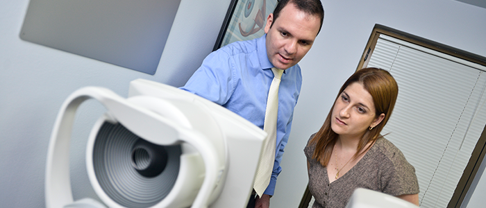
Complete ophthalmological exam
The complete ophthalmological examination includes visual acuity study, refraction (measure of glasses), taking intraocular pressure, and microscopic review of the eyelids, iris, conjunctiva, cornea and crystalline lens. Subsequently mydriasis (dilated) pupil and eye fundus review to assess the retina and optic nerve.
According to the findings some complementary tests for accurate diagnosis may be necessary.