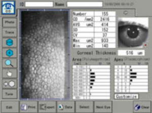
Corneal Topography
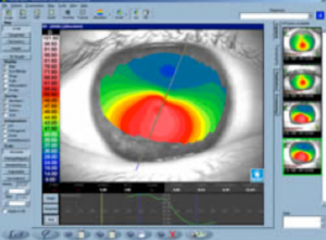 Corneal topography is a completely computerized test used to microscopically map the shape and the thickness of the cornea. This makes possible to detect diseases still in very early stages of its evolution, treatment planning, and allows us to identify patients who are candidates for laser refractive surgery or not.
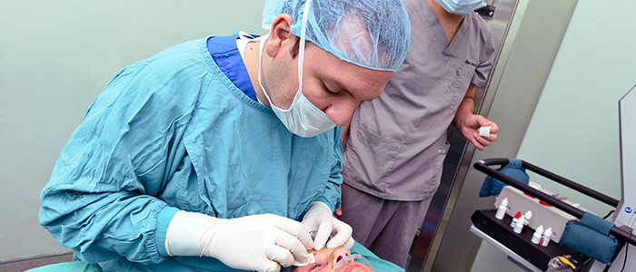
In disfiguring corneal diseases such as keratoconus, keratoglobus and pellucid marginal degeneration is mandatory to perform a topography to characterize and know their status, this will also provide us with the necessary information to perform procedures such as placement of INTACS stromal segments or crosslinking. After the treatment allows testing detailed changes and making other therapeutic necessary decisions.
For laser refractive surgery (myopia, hyperopia or astigmatism) topography provides the information necessary first to determine whether the patient is a suitable candidate and will have good results with treatment, second these data are needed to program the laser device and treatment planning, and third after surgery allows us to evaluate the results objectively.
After the corneal transplant, topography is needed to plan the most appropriate removal of stitches to get the best final visual outcome. In some cases of cataract surgery is very useful to calculate the power of the lens to be implanted in the eye during the operation and that is the one that will regain the patients vision properly. The more accurate the calculation is the best visual outcome the procedure will have.
The corneal topographer Galilei gives us additional information such as the distances and spaces within the eye and the hardness of the cataract prior to surgery. While our surveyor Magellan provides us extra information for contact lens fitting and surgical treatment of keratoconus.
Biometrics
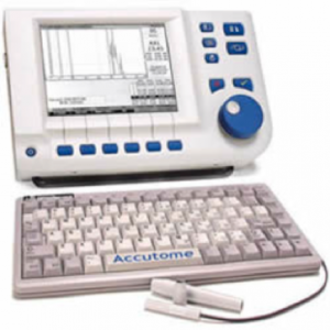
It is known as biometrics to the measure of the internal size of the eye, with these measures is possible to calculate the power of the lens placed inside the eye either during cataract surgery or refractive surgery with phakic lens.
For this examination a high accuracy of these measurements is essential, since this depends on the visual quality of the patient after the operation. Currently in the world gold standard (best method) taking these measurements is by using ultrasound especially designed for this purpose and using immersion techniques. Currently we have the Biometer Quantel Compact Touch that gives us personalized information of high precision and guarantees the best visual outcome possible in each patient.
Specular Microscopy
 Before any surgical procedure that involves working within the eye or the cornea is necessary to evaluate the ability of this to maintain transparency after the procedure. This transparency is essential for good vision with the operated eye and directly depends on the cell population (endothelium) of the posterior part of the cornea (internal). The better the endothelium, better and faster will be the recovery of the patient after surgery and lower the risk of having to perform in the short or medium term secondary corneal transplant complications.
Specular microscopy is a test that assesses the quantity and quality of these cells, providing the information necessary to present a customized surgical plan and further develop an appropriate visual prognosis for each patient.
We have the latest technology in this field represented by Tomey EM 3000 specular microscope.
High resolution OCT
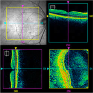
Currently we offer consistent high-resolution scanner Zeiss Cirrus, which has a spectral latest technology and is unique in the country.
The optical coherence tomography (OCT) allows to analyze the anterior and posterior segments of the eye. In the first segment can be observed in detail the cornea, iris and half of the crystalline lens, and it enables to study iris lesions such as cysts; cataract severity, and moreover, in refractive surgery, to evaluate the thickness of the cornea. In the posterior segment the equipment examines the retina and its pathologies, therefore can detect diseases such as macular degeneration and also allows studies of optic nerve and nerve fibers in the diagnosis of glaucoma, even early.
It is very useful for the diagnosis of glaucoma because it can accurately measure the amount of nerve fibers in the optic nerve, which decrease in diseases like glaucoma, such devices even say where the fibers are decreased and in what quantity. The equipment also effectively allows patients to track their ability to show minimum changes that are not seen clinically.
In general, it allows to see such small defects that might be overlooked in an standard OCT resolution. It is a diagnostic tool that complements the established diagnosis and also allows us to analyze the evolution of patients and determine whether they have improved or are getting worse and are responding to treatment.
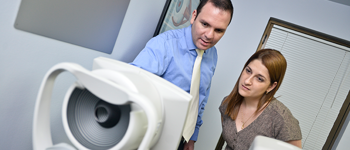
Ocular Aberrometry Total with OPD 3
It is the latest Nidek ophthalmological innovation, company leader in ophthalmology in the world. Permits to get rich and accurate information on the refractive status of the patients and its software helps get the ideal results in various procedures ranging from the adaptation of a contact lens, to cataract surgery or refractive laser surgery.
In one device combines a topographer, an aberrometer, a keratometer autorefractometer and pupillometer, capturing and analyzing thousands of data in a few seconds to give the doctor as much information about the eye, the visual quality and the necessary adjustments to optimal surgical result.