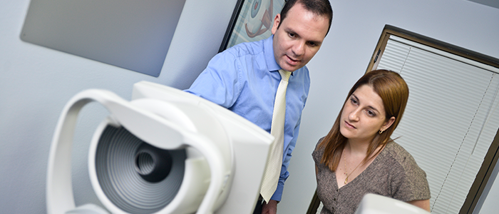
Micro-incision cataract surgery
The eye has an inside lens called crystalline, which under normal conditions is completely transparent and adapts to allow focus images at all distances. Over time the lens undergoes a process of degeneration, which first manifestation is the loss of the ability to focus up close (presbyopia) and subsequently loses transparency and produces blurred and distorted vision. This loss of the transparency is what is known as a cataract, and is progressive over time and may even lead to blindness.
Modern cataract surgery, such as that made by us, allows the restoration of good quality of vision and depending on the lens used in the operation, also allows the restoration of the ability to focus on near and far.
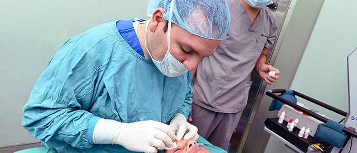
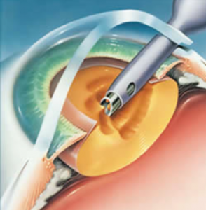
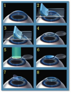
This surgery is performed using an ultrasound machine specially designed called phacoemulsificator. This device allows virtually undoing the cataract in the eye and placing a lens that replaced it, and allows the patient to return to see clearly. This entire procedure is performed through an incision of only 2 mm, made in the clear part of the eye (cornea) and using only anesthetic drops, without injections or general anesthesia and reducing surgical risks to a minimum, allowing performing surgery in elderly or even chronic diseases.
Currently, Dr. Centeno uses the Infinity phacoemulsifier of Alcon Laboratories, this is the most advanced and secure worldwide. The Ozil© technology of this device has revolutionized cataract surgery, shortening the time of surgery and minimizing the risk of complications to unprecedented levels. This coupled with his facochop surgical technique which offers surgical success in at least 98% of the cases operated.
Advanced Keratoconus Surgery (INTACS – Crosslinking)
 The cornea is the transparent lens located on the front of the eye, just ahead of the pupil and is essential for good vision because provides two-thirds parts of the focusing power of the eye. In special situations progressive deformation processes of the cornea is produced, which leads to the generation of high myopia and astigmatism, this leads to progressive and severe vision impairment in a large proportion of cases.
These corneal disfiguring disease are known as ectasia and include keratoconus, keratoglobus and pellucid marginal degeneration. Formerly for these cases there were no much treatment options, hard contact lenses were used, known for its difficult adjustment and multiple constraints for use. In more advanced cases was indicated corneal transplant surgery, highly complex surgery, with subsequent risk of rejection or complications and unsatisfactory refractive results in much cases.
Fortunately there have been great advances in the treatment of these diseases in recent years and already have in our clinic the possibility of corneal crosslinking and placing INTACS intra-corneal segments.
The corneal crosslinking is a minimally invasive procedure that allows, through chemical reactions induced in the cornea, strengthening fibers that compose it, getting to stop the disease in a very high percentage of cases and even decrease the degree of ectasia in some of them. All this is achieved by instillation and impregnation of the corneal tissue with special vitamins, that when exposed to concentrated ultraviolet light react forming bonds between the fibers that compose the cornea and that stronger arresting the progress of the deformation.
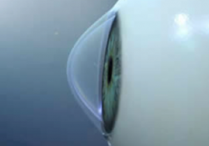
INTACS segments are semicircular small fragments of a special clear plastic; these are placed in the cornea, about the center. They exert a mechanical force that restores the original form, significantly reducing myopia and astigmatism and improving the visual quality of the operated person. While there are other options of segments in the market, the Intacs are the only approved to use in the U.S. by the FDA, as they have proven through countless clinical studies its effectiveness and safety, making them the best choice today in the world for correction of keratoconus. Dr. Centeno is in Costa Rica one of the only three certified surgeons for placement of Intacs. For more information visit the page www.intacsforkeratoconus.com.
In our clinic we offer the services of corneal crosslinking, cornea transplant and placement of Intacs segments. We also have the option of last generation contact lenses, hybrids and three curves for handling ectasias in people who cannot operate and are looking for better options than traditional hard contact lenses.
Laser refractive surgery
 Refractive surgery allows correction of the eye’s focusing defects such as myopia, hyperopia and astigmatism. These refraction vices consist in when light entering the eye is not properly addressed to the retina; this results in blurred vision, eyestrain, difficulty in reading, headaches and even dizziness.
To correct these problems eyeglasses or contact lenses are used, but they generate a dependency in the sense that for the proper functionality of the individual are strictly necessary. And when for some reason they do not have them, serious limitations in their activities occur.
Modern surgery can rid of these visual aids, improving the quality of life and even the aesthetics of the people who undergo it. There are several options on the market; the best known is the laser, which has already proven its effectiveness and security for many years in the correction of refractive errors. However, for cases of patients who for some reason are not candidates for this procedure, there are very effective options, including phakic lenses (placed inside the eye) and phaco-refractive surgery (replacement with an artificial lens), which allows to virtually correct any defect in focus.
In our clinic we offer services in refractive surgery with very high levels of satisfaction and safety in all patients using the latest technology in excimer laser (LASIK), phakic lenses and phaco refractive surgery.
Corneal Transplant
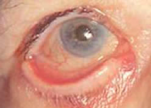
 Corneal transplantation or penetrating keratoplasty as it is also known, is a surgical technic in which the diseased corneal tissue is replaced throughout its thickness, for a healthy cornea from a donor. It is a procedure that has been practiced in our country for many years with excellent results.
Candidates for this surgery include people with diseases of the cornea untreatable by other means as very advanced keratoconus, people with severe and profound corneal scars that do not allow them to see well, severe corneal infections, permanent decompositions of the cornea after complicated cataract surgery or in case of severe dystrophic diseases of the cornea with a high degree of visual alteration.
This is a highly complex surgery both during surgery, as well as in the recovery period. In this, proper medication and regular checkups are essential for a correct final result.
Glaucoma Surgery
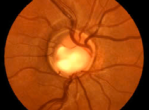 There are patients in whom medical treatment (drops and tablets) is insufficient to ensure adequate control of the glaucomatous disease. For them the best option is surgery, which although complex, offers excellent results, lowering intraocular pressure and stopping the process of damage to the optic nerve.
There are several different types of glaucoma surgery, most focus on encouraging the exit of fluid from the eye to relieve the pressure. In some cases specially designed valves implanted or laser application in the filtration zones of the eye is necessary.
In our clinic we offer trabeculectomy surgeries, Ahmed valve implant, laser trabeculoplasty and iridotomy. In advanced cases of refractory we offer cyclocryotherapy (freeze surgery) partial or total to control of blind painful eye.
Pterygium surgery
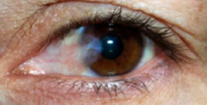 A pterygium is a degeneration of the conjunctiva (web that covers the white part of the eye) that is produced mainly by sustained exposure to sunlight. That is why is frequently seen in fishermen, outdoor workers or people whose lifestyles are exposed to ultraviolet light. In our country and in other tropical regions is very common in the population.
It appears as a small piece of cloth with blood vessels that give a reddish or inflamed appearance, most often located on the side of the nose, born in the white of the eye to insert the other end into the colored part (precisely in the cornea). As time goes on it tends to grow, invading more towards to the center of the cornea, causing difficulty seeing when it is advanced and also begins to produce symptoms such as burning, red eye, tearing and grittiness.
While early stage symptomatic relief is achieved with eye drops, in advanced stages surgery to the final resolution of the problem is necessary. Unfortunately there is a significant percentage of between 5 and 10% of pterygia that come out again and this percentage may be higher (up to 30 or 40%) according to the surgical technique used to resect. That is why we recommend performing surgery associating in the same procedure a graft or conjunctive transplant taken from the same eye, which minimizes the risk of the disease returning.
The conjunctive graft is a sophisticated technique and requires the placement of sutures, which are removed between 10 and 15 days after the surgery. In addition the use of anti-inflammatory eye drops, lubricants and antibiotics are required to prevent complications such as infections.
Eyelid Surgery
 The most frequent eyelid abnormalities include entropion (eyelid turned inward), ectropion (eyelid turned out), trichiasis (eyelashes flipped inward), chalazia and other tumors.
These problems tend to be annoying and cause further impairment of the aesthetic eye. In the case of tumors diagnose is important to define whether they are malignant and remove it promptly to avoid serious complications.
In our clinic we offer surgery to correct all these eyelid problems timely, safely and in the best hands.
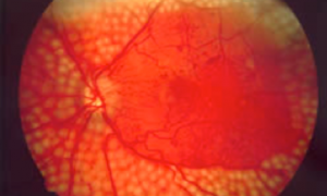
Laser treatment of diabetic retinopathy
 Diabetes causes serious eye injuries including cataracts, bleeding within the eye, swelling (edema) and retinal detachment, and even glaucoma (increased eye pressure). The appearance of some degree of injury is the rule in all diabetic patients, however adequate control of blood sugar levels slows and reduces the risk of visual loss.
In every person suffering from diabetes an eye full valuation every year in order to detect early-stage lesions is needed, while still not causing vision disturbances and thus treat them at an early stage before they cause irreversible damage the eye.
Diabetic cataract is surgically treated replacing the opaque crystalline lens with an artificial lens that allows the recovery of vision. When damage is retinal laser treatment is necessary to prevent the progression of it, and to seek recovery of the patient’s visual quality.
This laser treatment consists in small burns, which are conducted in a very controlled manner and are located in affected eye areas. In this manner the diseased tissue is removed and the internal eye oxygenation is improved. The progress of the disease is controlled and the risk of vision loss or severe complications is controlled.
The procedure is performed under local anesthesia only in drops and requires no cuts or wounds in the eye, it is only necessary to dilate the pupil. It takes about 15 to 20 minutes per treatment session. The intensity of it and the number of sessions depends on the degree of ocular damage at that time.
After application of the laser regular monitoring is necessary every 3 to 6 months to evaluate illness response to laser. In some cases it is necessary to repeat the procedure until the desired results are achieved.
When there is dense cataract that does not allow a correct visualization of the eye fundus, early surgery is necessary to perform an adequate laser treatment.
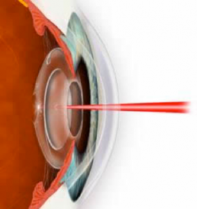
Laser cleaning of intraocular lens
 In people previously operated of cataracts, secondary to scarring process after eye surgery, opacity in the tissue surrounding the intraocular lens may be produce (secondary cataract). This can cause a decrease in quality of vision causing difficulty in reading, see details, glare or sensation of seeing through fog or mist.
Currently a non-invasive microsurgical process resolves this, conducted with a special laser disruptor, which has the ability to remove the opaque eye tissue from outside, literally without pain or discomfort, even without anesthesia. It is an extremely fast (about 5 minutes or less), safe and highly effective procedure, improving the visual quality almost immediately. It must be executed prior to a complete eye examination by a qualified ophthalmologist.
Surgical correction of presbyopia with Restor lenses or laser
With aging eye suffers deterioration of its close focus system, manifested in a progressive difficulty of seeing objects or read at short distance. This condition is known as presbyopia.
Presbyopia is a physiological process, i.e., it is not a disease. It is produced because the crystalline lens (lens inside the eye) becomes more rigid time and loses its ability to change shape to properly focus light coming from nearby sources, whether a book, newspaper, computer or any object. This forces the use of magnifying glasses for these activities.
Like any other degenerative condition is impossible to restore the youthful functioning to the eye with medication only, however, with the right technology, you can restore the ability to close focus and therefore avoid the perennial need for glasses.
Surgical correction of presbyopia may be performed intervening in one of two structures of the eye, or both sequentially. These include the cornea and crystalline lens.
In the case of the cornea we can, by using laser light, manipulate its shape and thus and so its focusing power to compensate for that lost by the aging eye. This can be achieved in two ways; the first is called Presbylasik in which a series of lenses to the cornea, some to near vision and others for far, are literally sculpted. The other possibility is mono-vision, which involves adjusting the focusing power of the eye to close or far vision, allowing the person to see at all distances, most patients adapt successfully, however it is previously required tests to verify the feasibility of the method in a personalized way.
When intervening crystalline what is sought is to replace the defective or degenerate structure for an artificial one with the ability to provide vision at all distances. This is achieved by a similar cataract surgery, and has the additional advantage that the disease will never develop because once the crystalline lens is removed there is not a substrate for the development of opacity that characterizes the cataract. The artificial lens implanted is known as Acrysof Restor and is the latest development so far in the treatment of presbyopia.
In our clinic we offer these new treatment options, after a complete evaluation and interview to define your own eye features and personal needs for your, allowing us to offer the best surgery for your particular situation.